Author Affiliations
Abstract
1 School of Electronic and Optical Engineering, Nanjing University of Science and Technology, Nanjing 210094, China
2 Smart Computational Imaging Laboratory (SCILab), Nanjing University of Science and Technology, Nanjing 210094, China
3 Department of Electrical and Computer Engineering, Boston University, Boston, Massachusetts 02215, USA
4 e-mail: leitian@bu.edu
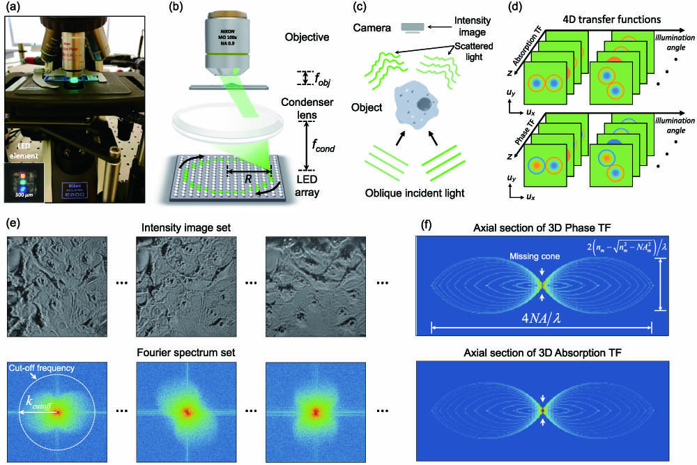
We propose label-free and motion-free resolution-enhanced intensity diffraction tomography (reIDT) recovering the 3D complex refractive index distribution of an object. By combining an annular illumination strategy with a high numerical aperture (NA) condenser, we achieve near-diffraction-limited lateral resolution of 346 nm and axial resolution of 1.2 μm over volume. Our annular pattern matches the system’s maximum NA to reduce the data requirement to 48 intensity frames. The reIDT system is directly built on a standard commercial microscope with a simple LED array source and condenser lens adds-on, and promises broad applications for natural biological imaging with minimal hardware modifications. To test the capabilities of our technique, we present the 3D complex refractive index reconstructions on an absorptive USAF resolution target and Henrietta Lacks (HeLa) and HT29 human cancer cells. Our work provides an important step in intensity-based diffraction tomography toward high-resolution imaging applications.
Photonics Research
2020, 8(12): 12001818
Author Affiliations
Abstract
1 Nanjing University of Science and Technology, School of Electronic and Optical Engineering, Nanjing, Jiangsu, China
2 Boston University, Department of Electrical and Computer Engineering, Boston, Massachusetts, United States
We demonstrate a label-free, scan-free intensity diffraction tomography technique utilizing annular illumination (aIDT) to rapidly characterize large-volume three-dimensional (3-D) refractive index distributions in vitro. By optimally matching the illumination geometry to the microscope pupil, our technique reduces the data requirement by 60 times to achieve high-speed 10-Hz volume rates. Using eight intensity images, we recover volumes of ~350 μm × 100 μm × 20 μm, with near diffraction-limited lateral resolution of ~ 487 nm and axial resolution of ~ 3.4 μm. The attained large volume rate and high-resolution enable 3-D quantitative phase imaging of complex living biological samples across multiple length scales. We demonstrate aIDT’s capabilities on unicellular diatom microalgae, epithelial buccal cell clusters with native bacteria, and live Caenorhabditis elegans specimens. Within these samples, we recover macroscale cellular structures, subcellular organelles, and dynamic micro-organism tissues with minimal motion artifacts. Quantifying such features has significant utility in oncology, immunology, and cellular pathophysiology, where these morphological features are evaluated for changes in the presence of disease, parasites, and new drug treatments. Finally, we simulate the aIDT system to highlight the accuracy and sensitivity of the proposed technique. aIDT shows promise as a powerful high-speed, label-free computational microscopy approach for applications where natural imaging is required to evaluate environmental effects on a sample in real time.
computational microscopy three-dimensional imaging diffraction tomography phase retrieval Advanced Photonics
2019, 1(6): 066004


